A team of researchers from the United States and Australia have found a link between the genes that regulate iron in the body and brain structure. More importantly, they found the link to be genetic – using twins.
A team of researchers from the United States and Australia have found a link between the genes that regulate iron in the body and brain structure. More importantly, they found the link to be genetic – using twins.
“If you use twins, you can basically implicate DNA,” Paul Thompson, study co-author and Professor of Neurology at UCLA, told Science-Fare.com. “You can find out if there’s something at the genetic level that causes iron to be linked to brain structure or some genes that affect both of them – and that’s what we did.”
After finding an association, the researchers did what’s called a cross-twin, cross-trait analysis. Using identical twins, they measured the iron levels of twin A and compared it to the brain structure in twin B.
If the association is genetic, the information should allow them to predict the brain structure of twin A more often than not.
Then, they repeated the experiment with fraternal twins. Because they don’t share exact copies of DNA like identical twins, the prediction should only be right, at most, half the time – like the odds of guessing heads on a coin.
“The fraternal twins are a control,” Thompson said. “Siblings are alike because they’re brought up by the same parents and share similar life experiences in childhood.”
Comparing the predictions in identical twins and fraternal ones allows researchers to control those nurture factors and directly implicate nature. If the prediction rates are better in identical twins than fraternal ones, the link is genetic – and they were.
“We now knew the same sets of genes were implicated in iron regulation and brain structure,” Thompson said.
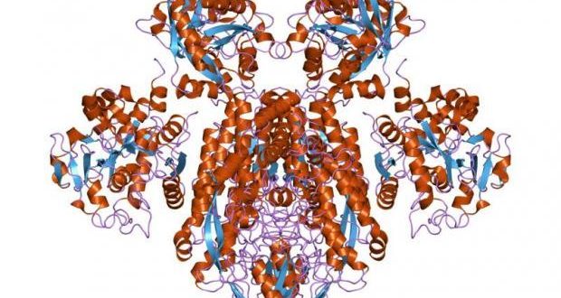
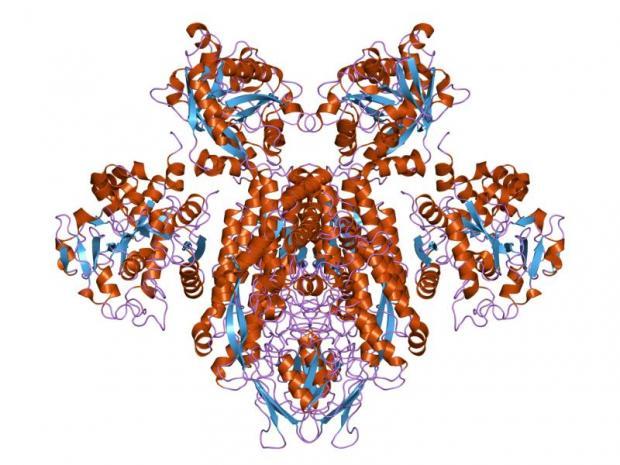
In the blood, researchers say iron is tightly regulated by the protein transferrin. It acts like a sponge and stores iron when it’s in excess. When iron levels are low, the amount of transferrin in the blood is increased.
“Transferrin levels don’t vary a terrific amount,” Thompson said. “So, it’s more effective for measuring iron levels than by measuring iron itself.”
In the brain, iron is regulated by specialized cells called oligodendrocytes. They’re responsible for insulating the nerves and iron has been shown to support myelin formation – the nerves protective coat.
In order to get into the brain, iron from the blood is transported to the blood-brain barrier by a transferrin molecule. There, it binds to its receptor before the iron alone is shuttled across the membrane.
Research has shown another gene, HFE, is important for facilitating that transfer. Defects in the HFE gene lead to a condition known as hemochromatosis.
“Approximately one per cent of the population has the defect in the HFE gene that’s extremely serious,” Thompson said. “Iron overloads in the brain and can be very toxic.”
“Ten per cent of the population carries a less serious mutation in the gene just seems to give brain differences,” he added. Iron loading still occurs, but not at a noticeable rate.
When the researchers looked at these patients, they had less transferrin compared those without the mutation and had an increased amount of white matter in the brain – more evidence of a link between brain structure and iron regulation.
The researchers say this discovery could help them understand the underlying mechanisms that affect cognition, neural development and neurodegeneration.
The research was published in the latest issue of the Journal, Proceedings of the National Academy of Sciences.
 Science Fare Media Science News – Upgraded


