Related Articles
Science Fare Media’s Managing Editor, Lee Flohr, went one-on-one with Dr. Robert Lefkowitz to talk about the science that resulted in a call from Stockholm.
Two medical doctors from the United States have been awarded the 2012 Nobel Prize in Chemistry for helping understand how that science drives the biology of an important bunch of proteins, known as G-protein-coupled receptors.
Stitched into the fabric of the cell a hallmark seven times, they help our brain make sense of our senses and allow our cells to communicate with each other.
Science Fare Media’s Managing Editor, Lee Flohr, spoke one-on-one with Duke University’s Dr. Robert Lefkowitz – who shared this year’s Nobel Prize with Dr. Brian Kobilka, a former post-doc in his lab – to learn more about the experiments that spurred a science and saved him from Vietnam’s front line.
Before the GPCRs could even be studied, the chemistry to study them had to be developed. It was Dr. Lefkowitz who designed radiolabeled compounds that would allow researchers to monitor their interaction with the cell.
They subsequently found the hormone or neurotransmitter – like adrenaline or serotonin – attaches to the receptor on the outside of the cell and triggers an independent cascade inside.
Since then, roughly a thousand GPCRs have been discovered in humans, several have even been imaged directly and a respectable amount, are targets for important pharmaceutical drugs.
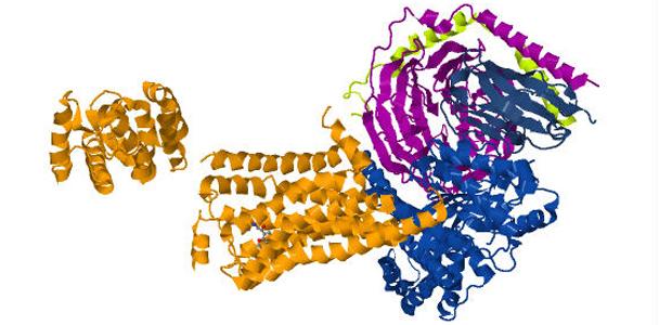
Here’s a guide for understanding the structure pictured above – and mentioned by Dr. Lefkowitz in our interview. It’s a cartoon to help make it easier to understand all the parts.
The orange bunch in the centre of the picture is the actual receptor – it’s hard to look in 2D, but you can count seven spirals that span the membrane and help keep it anchored there.
The right side of the cartoon is how the GPCR looks inside the cell. The lighter blue, purple and green portions are the G-protein that’s coupled to the receptor – hence the name – and the darker blue portion is a remnant of a tool needed to get the job done.
The second orange portion on the left side is also a remnant. It’s also on the outside of the cell where the receptor binds the chemical compound – which can be seen in the left side if you look closely – and triggers the cascade, without allowing the compound to actually enter the cell.
The American Chemical Society has made several of their research papers available, here.